
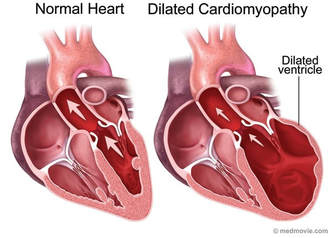
The normal heart is shown on the left compared to a heart with dilated cardiomyopathy on the right. Note the increased dimensions of the left ventricle.
A cardiomyopathy is a descriptive condition where the muscles of the heart is abnormal. There are four types of cardiomyopathies: “hypertrophic”, “dilated”, “restrictive” and “right ventricular”. While dilated cardiomyopathy, or DCM, has typically been recognized by its structure i.e., thinning and stretching of the heart muscle, the electrical function of the heart can also become adversely affected. When the heart chambers dilate, the heart muscle doesn’t contract normally. Also, the heart can’t pump blood very well. Over time, the heart becomes weaker and heart failure can occur. Dilated cardiomyopathy also can lead to heart valve problems, arrhythmias (irregular heartbeats) and blood clots in the heart.
The main feature of dilated cardiomyopathy is an excessive stretching of the heart muscle. The disease often starts in the left ventricle, the heart’s main pumping chamber. The heart muscle begins to dilate (stretch and become thinner). This causes the inside of the chamber to enlarge. When the heart chambers dilate, the heart muscle doesn’t contract normally. Also, the heart can’t pump blood very well and becomes unable to supply the body with enough blood. The problem often spreads to the right ventricle and then to the atria as the disease gets worse. Over time, the heart becomes weaker and heart failure can occur. Dilated cardiomyopathy also can lead to heart valve problems, arrhythmias (irregular heartbeats) and blood clots in the heart.
The cause of dilated cardiomyopathy often isn’t known. Certain diseases, conditions and substances also can cause the disease, such as poor genetics, congenital (from birth) heart defects, infections, parasites, lead toxicity, heart attack or high blood pressure.
Normally, the heart is set up to process deoxygenated blood and make it oxygenated to send to the tissues of the body. There are four chambers of the heart and four one way valves to continue pumping blood in this uni-directional process. The right atrium receives blood from the body which is poorly oxygenated, sends it to the right ventricle, where it is then transferred to the lungs. Blood becomes oxygenated and carbon dioxide is expelled. From there, the blood goes to the left atrium, then to the left ventricle, which pumps it to the body. This cycle is continuously repeated as the heart beats. Every heartbeat results from an electrical signal starting at the right atrium and travels down through the heart through special conducting tissue which starts a contraction. In a heart with DCM, not only are the muscles of the heart stretched, but this change can sometimes interfere with this normal electrical signal. Some of the different types of electrical signal malfunctions are arrhythmias of differing severities. These electrical malfunctions can even cause a cardiac arrest.
When a heart is in DCM, the heart contracts (pumps blood) poorly because the muscle is over-stretched. The amount of blood which the heart can hold is therefore increased. Since it is so stretched, a large amount of blood is able to fill each chamber, but the heart’s ability to pump the blood from each chamber is reduced. Enlargement of the left ventricle may make it harder for your heart valves to close, causing a backward flow of blood and making your heart pump less effectively. This can sometimes result in backflow of the blood from one chamber to another (heart valve regurgitation), against the uni-directional valve. Changes in heart structure and changes in pressure on the heart’s chambers can lead to an abnormal heart rhythm (arrhythmia). Unfortunately, dilated cardiomyopathy can cause your heart to suddenly stop beating, or blood clots (emboli) because there is pooling of blood (stasis) in the left ventricle due to the large volume of fluid in that chamber. This can lead to blood clots, which may enter the bloodstream, cut off the blood supply to vital organs, and cause stroke, heart attack or damage to other organs. Arrhythmias can also cause blood clots.
Dilated Cardiomyopathy may be suspected because of symptoms, a murmur, or an abnormal radiograph. Because such symptoms could be caused by a large number of other conditions, an ultrasound is required to officially diagnose dilated cardiomyopathy. If an arrhythmia is heard, an ECG may be recommended as well. An ECG records the electrical signals from the heart and is performed by placing electrodes in the armpits and inner legs. In Dilated Cardiomyopathy the ECG usually shows an abnormal electrical signal due to muscle stretching. Some patients may only show minor changes or be normal on an ECG. Because ECG abnormalities are not specific to Dilated Cardiomyopathy and may be found in other heart conditions, an ECG may or may not be done depending on the stress level of your pet.
If there is a suspicion of DCM, there are a few recommended tests that a veterinarian may advise. First, your veterinarian may order blood tests which help with information about your pet’s heart and also may reveal if your pet has an infection, a metabolic disorder or toxins in the blood that can cause dilated cardiomyopathy. Next, a chest X-ray is important to check your pet’s heart for abnormalities in the structure or size and the lungs for any sign of fluid in or around your lungs.
To diagnose DCM, your veterinarian will want an ultrasound of the heart. The ultrasound of the heart is sometimes an echocardiogram, or an ECHO. An ECHO produces a picture of the heart, and if excessive stretching of the muscle can be seen, DCM is diagnosed. During the test the entire heart is measured including the walls, valves and other structures within the heart. Therefore ECHO provides a very thorough assessment of Dilated Cardiomyopathy, and it can differentiate DCM from other heart conditions that may be suspected.
Because DCM develops over a period of time, your pet may not be exhibiting any signs of DCM because the change in the heart began slowly. Signs and symptoms of DCM Dilated cardiomyopathy can appear along a spectrum of no symptoms, subtle symptoms or, in the more severe cases, congestive heart failure (CHF), which occurs when the heart is unable to pump blood well enough to meet the body tissue needs for oxygen and nutrients. Things that may indicate DCM in your pet, or a worsening of DCM can be shortness of breath (dyspnea), lethargy (tiredness), swelling of abdomen (ascites), swelling of limbs (edema), syncope (fainting), seizures (convulsions), or even sudden cardiac arrest (heart stops beating effectively requiring resuscitation). These symptoms can occur at any age and with any stage of cardiomyopathy, even if other more severe symptoms of congestive heart failure have not yet appeared.
Drug treatment or medication is given when some or all of the symptoms listed above are present. These medications must be given for the remainder of your pet’s life, and the heart will be evaluated by alternating bloodwork and radiographs every 3 months. Increasing, decreasing or discontinuing medication should only be done with consultation with your veterinarian. The choice of treatment will vary from individual to individual but the common drug groups used for treatment medications are listed below:
Drugs that have proved useful in the treatment of dilated cardiomyopathy and heart failure include:
Angiotensin-converting enzyme (ACE) inhibitors
ACE inhibitors are a type of drug that widens or dilates blood vessels (vasodilator) to lower blood pressure, improve blood flow and decrease the heart’s workload. ACE inhibitors may improve heart function. An example of this type of drug is enalapril.
Side effects include low blood pressure, low white blood cell count, and kidney or liver problems.
Angiotensin II receptor blockers
These drugs have many of the beneficial effects of ACE inhibitors and may be an alternative for people who can’t tolerate ACE inhibitors.
Beta blockers
A beta blocker slows your heart rate, reduces blood pressure and prevents some of the harmful effects of stress hormones, substances produced by your body that can worsen heart failure and trigger abnormal heart rhythms. Atenolol and propranolol are some examples of this medication type. Beta blockers may reduce signs and symptoms of heart failure and improve heart function.
Diuretics
Often called water pills, diuretics remove excess fluid and salt from your body. The drugs also decrease fluid in your lungs, so you can breathe more easily. Common diuretics include furosemide and spironolactone. Common side effects of diuretics include dehydration and abnormalities in the blood chemistries (particularly potassium loss), which is why it is important for regular blood rechecks when your pet has DCM.
Inotropic agents
Digoxin. This drug, also known as digitalis, strengthens your heart muscle contractions. It also tends to slow the heartbeat. Digoxin may reduce heart failure symptoms and improve your ability to be active.
Blood-thinning medications
Your veterinarian may prescribe drugs, such as warfarin (Coumadin), to help prevent blood clots. Side effects include excessive bruising or bleeding. This is not commonly prescribed
There are a variety of drug treatments currently used in Dilated Cardiomyopathy (DCM). The need for any treatment and choice of that treatment has to be made on an individual basis and may change in each patient over the years. It is very important to discuss your pet’s symptoms with the veterinarian and develop a treatment plan for your pet, as each patient with DCM is different. Medication should NEVER be stopped without first consulting with your veterinarian. While medications can be stopped some drugs can have a serious complication if stopped abruptly, please work with your veterinarian to manage all medications. You will be doing follow up examinations every 3 months.